X-Ray
Prior to X-Rays
A single radiographic session is not hazardous for the dog. We at Ark Veterinary Hospital sedate all patients to ensure a good quality safe imaging can take place.
Using a special ruler, the area of concern is measured in order to determine the thickness, and thus the length of exposure necessary to produce a good quality image. The dog is then positioned on the x-ray table in order to get an optimal view of the dog’s broken bone (or other area of concern).
X-ray Procedures
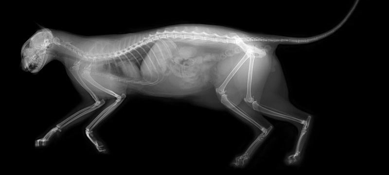
A plastic cassette containing the film is placed under the target area. The cassette prevents scratches or impurities from getting on the film and distorting the image produced. Veterinarians use different cassette sizes depending upon the size and shape of the affected area. The x-ray equipment is on a mechanical “arm” and is positioned over the area. The ray is triggered, creating images on the film in varying shades of gray based upon tissue density.
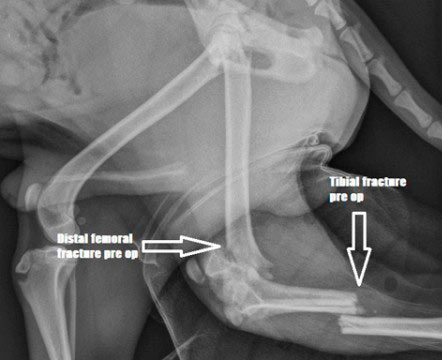
Repositioning the dog and taking additional film allows the veterinary staff to get multiple views. Multiple pictures reveal images of a break, tumor, or other health issue not visible in one image, but clear and distinct in another. The veterinarian reviews the films to diagnose the problem, and create a recommendation for treatment.


your pet since 1995
